Jack Cerchiara,'06 and Brendan Holsberry, 07
The Influenza A virus is a orthomyxovirus, and comprised of two primary structural protiens, Hemagglutinin (HA) and Neuraminidase (NA). It has been determined that Hemagglutinin is the primary protien responsible for binding to receptor sites on the cell membrane, allowing the virion to enter the cell (Subbarao 2000). Hemagglutinin is species specific binding protien that binds only to matched sialic acid receptors in host cells (Subbarao 2000). The molecule under study here is the HA protien extracted from the H1-human influenza strain.
The structure of Hemagglutinin is very similar between strains, and differs in only a few strucural difference. What makes this strain differ fromthe H5N1 influenza now posing a pandemic threat is the speficity of its binding regions, which will be the basis of this study. What makes this strain of influenza so dangerous is both its rapid ability to evolve and the fact that mammals, specifically humans contain to immune defences agaist this avian strain (Suarez 2000). In 1918, a strain of influenza A virus killed 20-40million people worldwide and the World Health Organization estimates nearly 7 million dead and 1 billion ill from an outbreak of the current H5N1 strain (Horimoto 2001).
Why look at Hemagglutinin?
Researchers have been able to determine that Hemagglutinin (HA) is a species specific binding protien that allows for the virus to bind to the cell membrane of host respiratory cells and propagate through cellular processes. By examining this process, medical researchers hope to determine a vaccine that may prevent this binding from occuring, thus preventing host infection.
Hemagglutinin is a trimer protien comprosed of a globular domain and a stem domain, divided along the longitudinal axis of the protien < >. As was stated, HA protien is made up of three monomers; HA1, HA2, and HA3 domains< >. Each of these monomers is comprised to two subdomains, <>, in the stem domain the two helix are bonded at Phe-88 < > to Phe-63 a clear difference between influenza strains, in the avian H5N1, it displays a inward facing Phenylalanine ring, while H1- human strains display an outward facing ring shown here. Along the longitiudinal axis, the protein is comprised of structural alpha-helicies and beta sheets < > are seen especially in forming the "bonding depression" in the globular region which will be discussed in the following section.
Note the beta-sheets are primarily present in the globular head < > where the binding region to sialic acid resides and the alpha helices < > make up the stem region of the HA monomers .
The hemagglutinin protien, as was previously stated, is a trinomer protien, primarily responsible for the binding of the Influenza A virion to cell surface receptors, membrane fusion and intracelluar infection, which is the first stage of viral infection. Hemaggluntin recognizes sialic acid components of cell-surface glycoprotiens and glycolipids (Gamblin et al., 2004; Ha et al., 2000). Hemagglutinin contains a shallow depression at its "head"< > which allows the sialic acid sequence to move into like a "lock and key." According to Ha et al. (2000), species specific sailic acid receptor analogs do not have a binding affect on the orientation of the sialic acid domain into the base of the hemagglutinin depression. One side of the sialic acid's pyranose ring faces the base of the binding depression. The axial carboxylate, acetamido nitrogen, and the 8- and 9-hydroxyl groups face the site and form bonds.
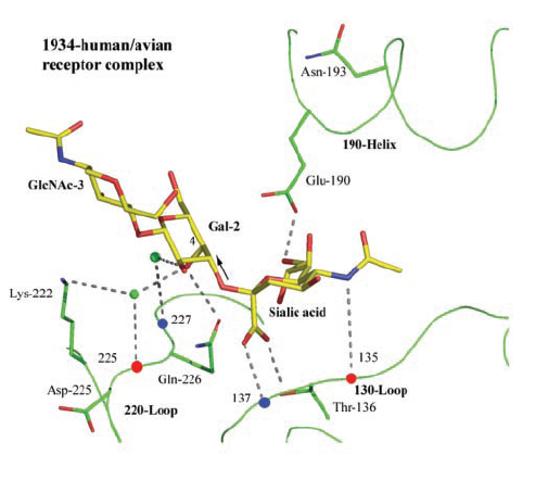
The binding depression surrounds the sialic acid domain with three primary regions of the hemagglutinin stucture. This region is comprised of a loop-helix-loop struture, which surrounds the sialic acid. The 130-loop, 190-helix and 220-loop structures form the trianglar opening into the beta-sheet depression.< > These three structures from the primary binding to the sialic acid and specific regions in their domains (Gamblin et al., 2004). The exact binding geometry differs between species speific hemagglutinin and cell surface protiens, however there are primary cites on the loop-helix-loop complexs that form speific interations allowing for the binding of the hemagglutinin and the subsequent cell infection. In human-avian receptor complexes the Glu-190 < > residue on the 190-helix forms a hydrogen bond to the 9-hydrxyl group. Thr-136 < > as well as amino-acids at residues 135 < > and 137 < >on the 130-loop form hydrogen bonds to the sialic acid's carboxylate. Also, Lys-222 < > and Gln-226 < >of the 220-loop form bonds with 8-hydroxyl group of the sialic acid (Gamblin et al., 2004) (Figure 1). Together this binding froms around the sialic acid domain of the cell surface glycoprotien or glycolipid in the HA depression, connecting each monomer to the sialic acid on the cell, initiating viral infection < >.
(Gamblin et al., 2004)
- Ya Ha, David J. Stevens, John J. Skehel, and Don C. Wiley. X-ray structures of H5 avian and H9 swine influenza virus hemagglutinins bound to avian and human receptor analogs. PNAS, Sep 2001; 98: 11181.
- S. J. Gamblin, L. F. Haire, R. J. Russell, D. J. Stevens,
B. Xiao, Y. Ha, N. Vasisht, D. A. Steinhauer, R. S. Daniels, A. Elliot, D. C.
Wiley, and J. J. Skehel
The Structure and Receptor Binding Properties of the 1918 Influenza
Hemagglutinin. Science, Mar 2004; 303: 1838 - 1842.
- Sauter, N. K., Hanson, J. E., Glick, G. D., Brown, J. H., Crowther, R. L., Park, S. J., Skehel, J. J., Wiley, D. C.: Binding of influenza virus hemagglutinin to analogs of its cell-surface receptor, sialic acid: analysis by proton nuclear magnetic resonance spectroscopy and X-ray crystallography. Biochemistry 31 pp. 9609 (1992)
- Subbarao, K.; Katz, J. Avian influenza viruses infecting humans. Cellular and Molecular Life Sciences. Vol: 57, Issue: 12, November, 2000, pp. 1770 - 1784
- Suarez, David L. Evolution of avian influenza viruses
Veterinary Microbiology, Vol: 74, Issue: 1-2, May 22, 2000 pp. 15-27
- Horimoto, Taisuke; Kawaoka, Yoshihiro. Pandemic Threat Posed by Avian Influenza A Viruses. Clinical Microbiolical Review., Jan 2001; Vol. 14: pg. 129 - 149.