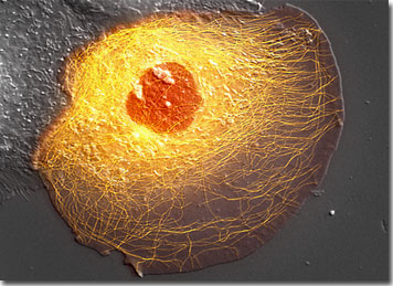
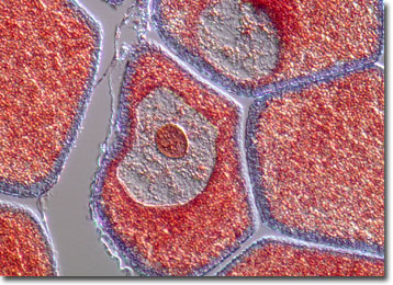
Drosophila larval hemisegment, 20x
Spring 2004
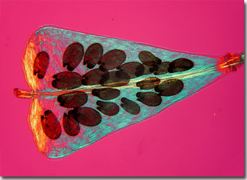
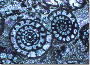
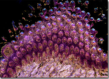
Capsella ursa-pastoris, 25x
Higley Room 214
Office Hours M,W 10-noon, T2:30-3:30
PBX 5398
| KENYON COLLEGE |
| BIO 346: Introduction to Microscopy and Image Analysis |
|
|
Spring 2004 |
|
| Fri @ 1:10pm, Higley 307 | ||
| Instructor: K.L. Edwards | ||
|
Higley Room 214 |
||
|
Office Hours M,W 10-noon, T2:30-3:30 |
||
|
PBX 5398 |
||
|
Text: Barbara
Foster. 1997. Optimizing Light Microscopy for Biological and Clinical
Laboratories. ASCLS. Kendell/Hunt Publishers. |
|
This laboratory is a practical course (not an experimental course) in light microscopy. It is designed to give you a theoretical basis in and a "taste" of the power and limitations of light microcopy as an investigative tool. Microscopy and image analysis protocols require time, diligence/patience, respect, and care. Some protocols may require weeks of work prior to observation. You must invoke patience and utilize great care to maximize the chances for successful outcomes. People proceed at different rates and returning to lab during the week to complete exercises is encouraged, rather than racing through them and garnering less. The pedagogy of the class is one of cooperative and collaborative learning. Discussion/queries/advice shared amongst us in the class is highly encouraged. I thank this and every class for informing changes that make this course a more effective and fruitful one. |
| Requirements: |
|
1. Attendance
2. Inquisitive and helpful attitude, diligence and perseverance.
3. Respect and care of the equipment (its all very expensive)
4. Project reports, both oral and annotated PowerPoint, as requested. (library and lab work)
5. 2 laboratory exam/practical. |
| Grade: |
Your grade is based on the quality of your lab practicals (20% each) and quality of your oral reports displayed in annotated PowerPoint (35-40%); class discussion and interaction (10-15%); and on your attitude, initiative and problem solving in lab, your laboratory methodology, and fulfilling obligations to the rest of class (10-15%). Missing a lab without permission or excuse from the Dean’s office will automatically drop your grade by 2/3 of a letter at the least. |
|
Human sperm, 1500x |
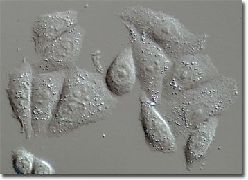
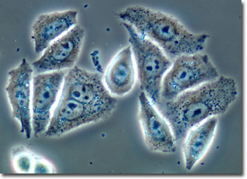
Mouse tongue
epithelia, 170x |
|
|
|
||
|
BIO3
46: Introduction to Microscopy and Image Analysis |
||
| LABORATORY SCHEDULE |
|
TEXT: Barbara Foster. 1997. Optimizing Light Microscopy for Biological and Clinical Laboratories. ASCLS. Kendell/Hunt Publishers. |
|
Thus the syllabus is not written in stone. We may not complete all; we may go further. We may head into other avenues of microscopic inquiry. Students and the instructor will present materials on aspects of microscopy and analysis throughout the course. |
|
Date |
Topic |
¨Text |
¨Lab ¨Links |
|
|
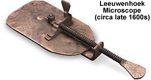
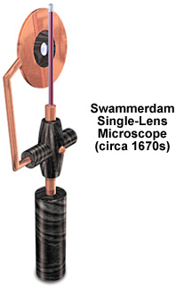
January 16
|
Early history of microscopy: The single lens microscope |
pp 141-142 |
Make single lens microscopes |
|
|
23 |
The dissecting and compound/inverted microscopes: Parts, use, care (Olympus CH2 video). Kohler alignment/illumination |
Intro Ch. 1
|
Observe organism’s cellular structure and its development with dissecting and compound microscopes. Submit drawings and description of organism. |
|
|
30 |
The nature of light and lenses Brightfield Student presentations on how the eye “sees”: visualization, retina structure and thresholds, optic nerve and brain decoding, color and contrast sensitivity |
Ch. 2 Ch. 8 Kohler handout |
Quantification: determination of actual magnification and making cellular measurements (handout) http://www.microscopyu.com/articles/ergonomics/ergointro.html eronomics of microscopy viewing http://www.microscopyu.com/articles/formulas/measurehome.html quantification http://www.microscopyu.com/tutorials/java/components/characteristicrays/index.html light and lenses http://micro.magnet.fsu.edu/primer/java/microscopy/immersion/index.html oil immersion |
|
|
February 6
|
Nature of light and lenses cont. Optimizing contrast techniques |
Chp 3 Chp 4
|
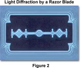
Handout on Airy disk http://micro.magnet.fsu.edu/primer/lightandcolor/diffractionintro.html http://micro.magnet.fsu.edu/primer/java/diffraction/basicdiffraction/index.html Diffraction and Airy disk http://micro.magnet.fsu.edu/primer/java/imageformation/rayleighdisks/index.html http://micro.magnet.fsu.edu/primer/anatomy/numaperture.html Airy disk http://www.microscopyu.com/articles/formulas/formulasindex.html concepts and formulas |
|
|
|
Optimizing contrast techniques Darkfield |
Ch. 5
|
Lab test http://micro.magnet.fsu.edu/primer/techniques/contrast.html contrast techniques http://micro.magnet.fsu.edu/primer/techniques/contrast.html darkfield http://micro.magnet.fsu.edu/primer/java/rheinberg/index.html Rheinberg http://micro.magnet.fsu.edu/primer/techniques/oblique/obliquehome.html oblique illumination |
|
|
20 |
Phase contrast microscopy
|
Ch. 6 |
http://micro.magnet.fsu.edu/primer/techniques/phasecontrast/phaseindex.html http://www.microscopyu.com/tutorials/java/phasecontrast/microscopealignment/index.html Phase Contrast http://micro.magnet.fsu.edu/primer/techniques/hoffman/hoffmanindex.html Hoffman Modulation Contrast |
|
|
27 |
Polarized light microscopy
|
Ch. 7: pp. 78-88 |
Convert a compound brightfield microscope to polarizing. Handout: polarized light and biological organization http://micro.magnet.fsu.edu/primer/techniques/polarized/polarizedhome.html http://www.microscopyu.com/articles/polarized/polarizedintro.html |
|
|
March 6-20 |
Spring Break |
|
|
|
|
20 |
Histology: the art of specimen preparation and slide making |
|
Handout |
|
|
|
Differential interference microscopy: DIC Histology cont. (sectioning with Spencer Microtome) |
Ch. 7: pp.89-95
|
http://micro.magnet.fsu.edu/primer/techniques/dic/dichome.html http://www.microscopyu.com/tutorials/java/dic/dicalignment/index.html
http://www.microscopyu.com/galleries/dicphasecontrast/index.html compares phase contrast and DIC images |
|
|
April 3 |
Fluorescence microscopy and image capture |
Ch. 7: pp. 72-78 Ch. 9 |
http://micro.magnet.fsu.edu/primer/techniques/fluorescence/fluorhome.html http://www.microscopyu.com/articles/fluorescence/filtercubes/filterindex.html http://www.microscopyu.com/tutorials/java/arclamp/index.html Fluorescence
http://www.microscopyu.com/articles/photomicrography/digital/index.html Digital imaging |
|
|
10 |
Limitations to light? Electron microscopy Histology cont. |
Ch. 10: pp 137-140 |
http://www.unl.edu/CMRAcfem/em.htm
|
|
|
17 |
Limitations to light microscopy? Viewing individual molecules in action with the light microscope. |
|
Lab test http://www.microscopyu.com/articles/general/innovations.html innovations http://www.microscopyu.com/articles/interferometry/twobeam.html interferometry...interference microscopy innovation
|
|
|
24 |
Deconvolution and Confocal microscopy: 3D imaging |
Ch. 10: pp. 133-134 |
Getting a stereo image from a compound microscope Resolving and quantifying in 3D (if system software is working) http://micro.magnet.fsu.edu/primer/techniques/confocal/index.html http://www.microscopyu.com/articles/confocal/index.html confocal http://micro.magnet.fsu.edu/primer/anatomy/stereohome.html http://www.microscopyu.com/articles/stereomicroscopy/index.html stereomicroscopy |
|
|
May 1
|
Student Presentations: Literature examples of how light microscopies are used to advance biological questions/research. |
|
|
|
| Click here to
access the quiz sets for this course. |
| Click here to access online reading assignments. |

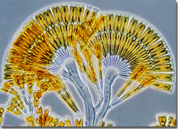
%2055x.jpg)