
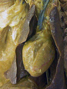
Figure 17. Ventral view of the heart lying in the mediastinal septum (left). Also associated with the septum
are fat bodies and the pink glandular thymus, which can be vaguely made out near upper center of the image. In
the image on the right the septum has been removed. The heart is still surrounded by the pericardial sac in the
picture on the right.