|
Right Internal View |
Left Internal View |
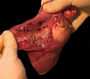
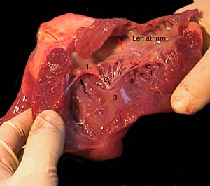
| 1 = Tricuspid Valve | 2 = Chordae tendinae | 1 = Bicuspid Valve | 2 = Chordae tendinae |
| 3 = Moderator Band | 4 = Inferior Vena Cava | 3 = Papillary Muscle | 4 = Trabeculae Carneae |
| 5 = Coronary Sinus | (Pulmonary Semilunar Valve) | Black Arrow - to Aorta | Red Arrow - to Pulmonary V. |