Anthrax Protective Antigen
Ansley Scott '02 and
Dawn Stancik '02
Biology 363
December 2001
Contents:
I. Introduction
Recently anthrax has caused much concern as a
public health threat because of its use as a bioterrorism weapon.
However, anthrax has been a concern for farmers since biblical times, especially
in agricultural regions like South America and Africa. Although it
is rare, anthrax has been found sporadically in cattle, sheep, and goats
throughout the midwest and the western United States after injestion or
inhalation of the Bacillus anthracis spores found in the soil.
Farmers rarely contract the disease and are usually only exposed to Cutaneous
anthrax through open wounds, whereby the bacteria can enter the skin.
Cutaneous anthrax is the form most encountered in naturally occurring cases
and is treatable; it is only deadly in about 20% of untreated cases (Anthrax
on the Farm )
. Inhalational anthrax
is very rare and usually fatal; after inhalation of the spores, cold symptoms
set in early and are abruptly followed by respiratory distress and death.
This form is the one used as a bioterrorist weapon. Anthrax can be
treated with antibiotics (ciprofloxacin) if diagnosed early enough, and
it is not a communicable disease. In bioterrorism, anthrax works
well because it is easy to prepare and disperse and can inflict sufficiently
severe disease to paralyze a city and perhaps a nation (Nature's
Anthrax Page
).
The Anthrax bacterium produces a toxin which
consists of the three proteins, the protective
antigen (PA), the lethal factor (LF), and
the oedema (OF) factor. These factors along with aa poly-D-glutamic
acid capsule are the major factors imparting virulence in B. anthracis.
The PA
protein works by forming a heptamer which binds to the surface of a host
cell and serves as a channel by which the oedema factor and the lethal
factor enter the cell (Heptamer).
Once in the cell, the PA protein is the common binding moiety of two toxins,
lethal and oedema, which are also composed of the lethal factor and the
edema factor proteins (Petosa, 1997).

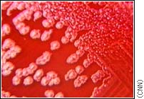
Bacillus
anthracis colonies
II. General Structure
The PA monomer
is predominantly made up of antiparallel b-sheets
and it consists of four functional domains (Petosa, 1997). Domain
I (residues 1-258)
,
the amino terminal domain, contains two calcium ions and the cleavage site
for proteases to activate the protein. The product of the cleavage
results in the creation of the amino terminal fragment a20 (Brossier, 1999).
Domain
II (residues
259-487)
is involved in the formation of the hexamer and has a flexible loop that aids
in membrane insertion. As of now, Domain
III (residues 488-595)
does not have a known function. Domain
IV (residues 596-735)
is needed for receptor binding ( Petosa, 1997).
III. Domain I and II
Domain I
PA is activated by proteolysis on the N-terminal
loop in Domain I. This causes the release of a 20 K fragment of Domain
I (PA20)
.
Loss of PA20 triggers
the heptamer formation of PA. This heptamer is soluble in water and
inserts itself into the host membrane (Heptamer)
(Petosa,
1997).
Domain II
The large loop in Domain II
(302-325)
is involved with the toxicity of anthrax and may be involved in the PA
channel interactions with the host. Residues
304,
306,
and 308
are located within the channel lumen (Petosa, 1997).
IV. Receptor Binding Region of Domain
IV
Domain IV was thought to contain two receptor binding
domains: the slarge loop(amino acid 704-723)
and the small
loop (amino acids 679-693)
.
It turns out that only the small loop
is involved in receptor binding (Varughese
et al., 1999).
In addition to the small loop,
residues at the end of the C-terminus aid in receptor binding. Deletions
in both the C-terminus and in the small loop
decrease anthrax toxicity by inhibiting binding. It has been shown
that two antibodies, 3B6 and 14B7, bind to the region between amino acids
671 and 721
and decrease toxicity by inhibiting the binding of PA to the receptor (Varughese
et
al., 1999)
.
V. Interactions With the Lethal Factor
The lethal factor
(LF)
is the major factor causing death in anthrax patients, but the mechanism
is not clearly understood. Internalization and translocation of the
lethal factor into the cytosol of Bacillus anthracis occurs when
the PA protein binds to a specific, yet undefined, cell surface receptor
(Pannifer, 2001). The highly specific LF enzyme has four domains
(I,
II,
III,
IV)
that have evolved through the gene duplication process, fusion and mutation
. After the LF has bound to the PA, the lethal toxin leads to an increase
in the permeability of Na+ and K+ ions after its
internalization and is followed by the hydrolysis of ATP. This inhibits
macromolecular synthesis involved with the immune response, and results
in cell death (Gupta et al., 2001). Interestingly, when the
PA and LF are injected in animals intravenously, they work in concert to
induce rapid death.
Domain III has a hydrophobic core (282-382).
It also contains a five-tandem repeat 101 amino acid sequence that seem
to have been created through a duplication event (282-382)
.
VI. References
1. Brossier, Fabien, Martine
Weber-Levy, Michele Mock, and Jean-Claude Sirard. 2000. Role of Toxin Functional
Domains in Anthrax Pathogenesis. Infection and Immunity. 68:1781-1786.
2. Gupta, Pankaj, Samer Singh, Ashutosh Tiwari,
Rajiv Bhat, and Rakesh Bhatnagar. 2001. Effect of pH on Stability of Anthrax
Lethal Factor: Correlation Between Denaturation and Activity.
Biochemical
and Biophysical Research Communications. 284: 568-573.
3. Pannifer, Andrew.
2001. Crystal Structure of the Anthrax Lethal Factor. Nature.
414:229-233.
4. Petosa, Carlo, R. John
Collier, Kurt R. Klimpel, Stephan H. leppla, and Rover C. Liddington. 1997.
Crystal Structure of the Anthrax Toxin Protective Antigen.
Nature. 385: 833-838.
5. Varughese, Mini, Avelino V. Teixeira, Shihui
Liu, and Stephen H. Leppla. 1999. Identification of a Receptor-Binding
Region Within Domain IV of the Protective Antigen Component of Anthrax
Toxin. Infection and Immunity. 1860-1865.