Murine Eukaryotic Translation Initiation Factor 4E bound to 7-methyl-GDP

Abby Lee '22 and Sumaya Ahmed '22


Contents:


I. Introduction

Murine eIF4E (Eukaryotic Translation Initiation Factor 4E) recognizes the 5’ 7-methyl-G(5’)ppp(5’)N cap at the end of messenger RNA and interacts with other proteins within the eIF4F complex to initiate translation. eIF4E acts as the first component that begins the recruitment of ribosomes to the 5’ cap structure, making this the rate limiting component in eukaryotic translation initiation because of its low abundance relative to the other factors and its ability to be bound to by inhibitor.

The eIF4F complex as a whole is involved in mRNA cap recognition, the unwinding of 5’-terminal secondary structure, and the recruitment of ribosomes. The complex is composed of eIF4E, eIF4G, a scaffold that binds together proteins in the complex, and eIF4A, an ATP dependent RNA helicase. Once the eIF4F complex is bound to the 5’ cap, the addition of eIF4B activates the helicase activity of eIF4A, leading to the unwinding of the secondary structure mRNA to allow the initiation codon to be accessible. Without eIF4E’s ability to recognize and bind to the 5’ cap of mRNA (here modeled with a 7-methyl-GDP) and to the conserved Trp-X-X-X-X-Leu-Leu (where X is any residue) of eIF4G, allowing the full assembly of all necessary components to stabilize the translational complex, eukaryotic translation would not occur as is.
eIF4Fcomplex

Figure 1. eIF4F complex containing eIF4E and eIF4G with eIF4A bound. (Papadopoulos et al, 2014)


II. General Structure

The three-dimensional structure of eIF4E is illustrated to the left. The protein is made up of amino acids 28-217 with one alpha/beta . This includes and alpha helices and an antiparallel beta sheet with . The three alpha helices are parallel to the strand direction and lie on top of the beta sheet. The mRNA cap is able to bind to the surface of the beta subunit, short alpha helix , and the loop connecting strand 1 and 2 (S1 and S2). Together, these components make the . eIF4E is phosphorylated at which induces a conformational change and increases the protein’s affinity for mRNA caps which in turn stimulates translation initiation. This activity can also be suppressed via binding proteins like 4E-BP.


III. mRNA Cap Binding

The 7-Methyl-GDP binds on the surface of eIF4E in the cap binding slot. The 5’ untranslated region of the mRNA exits over helix three (H3) and S4, S6, and S5. The N7 methylated GTP of the cap interacts with the S1-S2 and S3-S4 loops located between the side chains of conserved tryptophans Trp-56 and Trp-102. (All amino acids can be viewed within the structure ). Cap binding was reduced by at least 50% when substitutions were made that kept the pi-electron cloud, whereas other substitutions reduced binding all together, indicating the importance of the conserved sequence. eIF4E is a receptor for the cap by satisfying Watson-Crick base pairing.

Marcotrigiano et al (1997) divided the interactions between eIF4E and the mRNA cap into four classes as shown below.

I. Sandwiching of Alkylated Bases:
eIF4E recognizes bases that are alkylated. This occurs through the addition of an alkyl group, a functional group containing only carbon and hydrogen atoms, which facilitates bonding throughout mRNA cap binding. The 7-methyl guanine cap is “sandwiched” between the side chains of these alkylated bases. These interactions are all Van der Waals.

II. 7-Methyl Guanine Contacts:
Within the N7 methyl group there are oxygen atoms that bind to amino acids within the cap-binding slot, such as O6. Other atoms like N1 and N2 come between the two tryptophans involved in the alkylated base sandwiching, and bind with other amino acids, like Glu-103. These are largely hydrogen bonds and Van der Waals interactions.

III. Interactions between ribose and diphosphate:
The phosphate and ribose groups from the 5’ cap protrude towards the entrance of the cap-binding slot, but do not extend past the beta sheet. These groups interact with alkylated bases while being stabilized by hydrogen bonds and salt bridges.

IV. Water-Mediated Contacts:
Water-mediated contacts are shown below with two amino acids. The water molecule that is hydrogen bonded to the residue is highlighted in yellow. 


Class Amino Acids Interplanar Distance (A) Button
Sandwiching of Alkylated Bases

Trp-56 and Trp-102

3.5-3.6
7-methyl guanine contacts

Trp-102 and Glu-103 and Trp-166

2.7, 2.7-2.9, 3.7
Interactions between ribose and diphosphate

Trp-56 and Arg-157 and Lys-162

3.7, 3.0, 3.1
Water-mediated Contacts

Trp-166 and Arg-112

3.0, 2.8, 3.1 and 2.4, 3.2

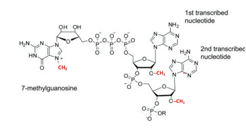
Shown below is the 7-methyl guanine structure within the 5' cap. Shown in red are the methyl groups on guanine at N7 and the ribose at positions 1 and 2. These modifications assist in the ability of eIF4E to recognize the 5' cap, thus promoting high affinity binding. This leads to the recruitment of eIF4F which signals for the initiation complex as shown in Fig. 1 to begin translation.
cap.png

Figure 2. Structure of the 7-methyl guanine of the 5'cap of mRNA. Methyl groups shown in red. (Cowling et al, 2010)


IV. eIF4G and eIF4E Complex Binding

Binding between eIF4G and eIF4E proteins is not as well known as the interactions between the mRNA cap and the eIF4E protein. It is known that there is a on the hydrophobic surface of all known eIF4E proteins that is thought to be important for eIF4G binding and interactions with 4E binding proteins. Translation initiation is thought to only proceed if eIF4E recognizes the sequence in  eIF4G (pictured in the button).

[Amino acid classes are labelled by color: conserved sequence, hydrophobic sidechains, polar uncharged sidechains, electronically charged sidechains, others.]

Marcotrigiano et al (1999) found this sequence in mammalian eIF4GI and eIF4GII to involve between hydrophobic residues Histidine-37, Valine-69, Tyrosine-624, and Phenylalanine-628. The eIF4E and eIF4G proteins share similar interactions with eIF4E and the mRNA cap, including hydrogen bonds, Van der Waals interactions, water mediated contacts, and eIF4G also utilizes salt bridges to contact residues. It is also known that these peptide residues do not hold a secondary structure unless eIF4E is present. The mechanisms and detail about binding between these proteins is very limited.


V. References

Marcotrigiano, J., Gingras, A.-C., Sonenberg, N., & Burley, S. (2000). Cocrystal Structure Of The Messenger Rna 5 Cap-Binding Protein (Eif4E) Bound To 7-Methyl-Gdp. doi: 10.2210/pdb1ej1/pdb

Marcotrigiano, J., Gingras, A.-C., Sonenberg, N., & Burley, S. K. (1999). Cap-Dependent Translation Initiation in Eukaryotes Is Regulated by a Molecular Mimic of eIF4G. Molecular Cell, 3(6), 707–716. doi: 10.1016/s1097-2765(01)80003-4

Rom, E., Kim, H. C., Gingras, A.-C., Marcotrigiano, J., Favre, D., Olsen, H., … Sonenberg, N. (1998). Cloning and Characterization of 4EHP, a Novel Mammalian eIF4E-related Cap-binding Protein. Journal of Biological Chemistry, 273(21), 13104–13109. doi: 10.1074/jbc.273.21.13104

Niedzwiecka, A., Marcotrigiano, J., Stepinski, J., Jankowska-Anyszka, M., Wyslouch-Cieszynska, A., Dadlez, M., … Stolarski, R. (2002). Biophysical Studies of eIF4E Cap-binding Protein: Recognition of mRNA 5? Cap Structure and Synthetic Fragments of eIF4G and 4E-BP1 Proteins. Journal of Molecular Biology, 319(3), 615–635. doi: 10.1016/s0022-2836(02)00328-5

Cowling, V.H. (2010). Regulation of mRNA cap methylation. The Biochemical journal 425, 295-302.

Shanmugam, R. (2014). Biochemical characterisation of tRNA-Asp methyltransferase Dnmt2 and its physiological significance.

Papadopoulos, E., et al. “The Co-Complex Structure of the Translation Initiation Factor eIF4E with the Inhibitor 4EGI-1 Reveals an Allosteric Mechanism for Dissociating eIF4G.” Proceedings of the Natural Sciences of America of the United States of America, 2014, doi:10.2210/pdb4tqc/pdb.

Back to Top