Back to: Biology 112 Syllabus
Back to: Biology 112: Evolution and Ecology Home Page
Back to: Kenyon College Biology Department
Assigned reading: Chapter 27 in text
Quiz this week? No. But you do have your first hour-long exam!
Study help:
Protists and the Dawn of the Eukarya
Protists are defined as all eukaryotes other than plants, fungi, and animals.
Most are unicellular, but many are multicellular or colonial.
Most are aquatic, both fresh and marine.
Some are parasitic.
Many are in moist organic matter or damp soil.
All other eukaryotes are presumed to have arisen from the protists.
How could eukaryotes evolve in the first place?
What changes had to occur to derive a modern, eukaryotic cell?
How can such a major change occur, and is there evidence for this?
What's inside a eukaryotic cell?
Are the cells of eukaryotes more complex than those of prokaryotes?
How are they different?
What kinds of evolutionary forces lead to such changes?
Read on!
Several steps had to occur for eukaryotic cells to become reality.
They include:
Androgenous model
The following diagram shows how infolding of the plasma membrane
adds surface area without increasing the cell's volume. One can easily
imagine how vesicles could form.
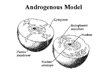
Diagram above from the University of Winnipeg Biology Dept. pages.
A fundamental concept of evolution is the belief in the natural progression
from the simple, to the more complex. For the evolution of the eukaryotic cell,
the predominating theory is known as the Endosymbiotic Theory. The
Endosymbiotic Theory of Eukaryote Evolution (Symbiotic Theory) was first
proposed by former Boston University Biologist Lynn
Margulis in the
1960's and officially in her 1981 book "Symbiosis in Cell Evolution".
Although
now accepted as a plausible theory, both she and her theory were ridiculed by
mainstream biologists for a number of years. Thanks to her persistence, biology
can now offer a plausible explanation for the evolution of eukaryotes. The theory
maintains that ancestors of eukaryotic cells were "symbiotic consortiums"
of
prokaryote cells with at least one and possibly more species (endosymbionts)
involved. In other words, perhaps oxygen breathing bacteria invaded an
anaerobic bacteria, and each performed mutually benefiting
functions. The bacteria would breathe for the anaerobic amoeba-like bacteria,
and the amoeba-like bacteria would navigate through new oxygen-rich waters in
search of food. This way, each of the organisms would be benefiting from their
symbiotic relationship as the waters and atmosphere of the Precambrian
changed.In support of this, notice that oxygen begins to accumulate between
the first fossil records of Prokaryotes and Eukaryotes.
Professor Kwang Jeon's Supporting Discovery Seem more like a creative
story than a plausible theory? Let's examine the case of Professor Kwang
Jeon of the University of Tennessee. In 1987, Professor Jeon noticed that his
collection of amoeba were developing a large number of dots. These large
number of dots turned out to be bacteria, which were quickly killing off Jeon's
collection. Jeon noted the least sick ones and began keeping records of their
progress. The least sick ones apparently were more resistant to the bacteria
since they survived and returned to their normal modes. However, some
40,000 of the invading bacteria were still present within each of the surviving
amoebas! Through transplanting experimentation, Jeon found that the nucleus
of the amoebas could not live without the once pathogenic bacteria. Jeon's
accidental discovery proves that it is possible for an organism to become
dependent on and a functional part of invading organisms. Rather than
eliminating competitors, evolution eliminated competition itself on the basis
of symbiotic relationships. (From J. Bond)
The figure below diagrams how this could occur.
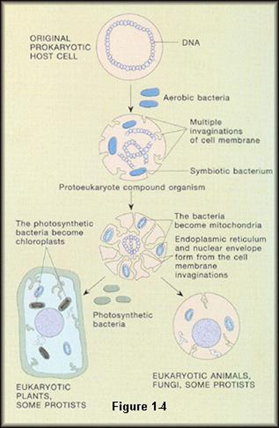
When did these events probably occur?
What do Protists look like?
(The four images and text in this section come from the University of
Calgary Dept. of Geology and Geophysics site about palynology and dinoflagellates.)
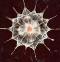
 There is a great deal of wonderful variability and diversity among
the protista. The two micrographs above show a couple of different dinoflagellates,
which are microscopic, (usually) unicellular, flagellated, often photosynthetic
protists, commonly regarded as "algae" (Division Dinoflagellata).
They are characterized by a transverse flagellum that encircles the body (often
in a groove known as the cingulum) and a longitudinal flagellum oriented perpendicular
to the transverse flagellum. This imparts a distinctive spiral to their swimming
There is a great deal of wonderful variability and diversity among
the protista. The two micrographs above show a couple of different dinoflagellates,
which are microscopic, (usually) unicellular, flagellated, often photosynthetic
protists, commonly regarded as "algae" (Division Dinoflagellata).
They are characterized by a transverse flagellum that encircles the body (often
in a groove known as the cingulum) and a longitudinal flagellum oriented perpendicular
to the transverse flagellum. This imparts a distinctive spiral to their swimming motion. Both flagella are inserted at the same point in the cell wall, by convention
defining the ventral surface. This point is usually slightly depressed, and
is termed the sulcus. In heterotrophic dinoflagellates (ones that eat other
organisms), this is the point where a conical feeding structure, the peduncle,
is projected in order to consume
motion. Both flagella are inserted at the same point in the cell wall, by convention
defining the ventral surface. This point is usually slightly depressed, and
is termed the sulcus. In heterotrophic dinoflagellates (ones that eat other
organisms), this is the point where a conical feeding structure, the peduncle,
is projected in order to consume  food.
Dinoflagellates possess a unique nuclear structure at some stage of their life
cycle - a dinokaryotic nucleus (as opposed to eukaryotic or prokaryotic), in
which the chromosomes are permanently condensed. The cell wall of many dinoflagellates
is divided into plates of cellulose ("armor") within amphiesmal vesicles,
known as a theca. These plates form a distinctive geometry/topology known as
tabulation, which is the main means for classification. Both heterotrophic (eat
other organisms) and autotrophic (photosynthetic) dinoflagellates are known.
Some are both. They form a significant part of primary planktonic production
in both oceans and lakes. Most dinoflagellates go through moderately complex
life cycles involving several steps, both sexual and asexual, motile and non-motile.
Some species form cysts composed of sporopollenin (an organic polymer), and
preserve as fossils. Often the tabulation of the cell wall is somehow expressed
in the shape and/or ornamentation of the cyst.
food.
Dinoflagellates possess a unique nuclear structure at some stage of their life
cycle - a dinokaryotic nucleus (as opposed to eukaryotic or prokaryotic), in
which the chromosomes are permanently condensed. The cell wall of many dinoflagellates
is divided into plates of cellulose ("armor") within amphiesmal vesicles,
known as a theca. These plates form a distinctive geometry/topology known as
tabulation, which is the main means for classification. Both heterotrophic (eat
other organisms) and autotrophic (photosynthetic) dinoflagellates are known.
Some are both. They form a significant part of primary planktonic production
in both oceans and lakes. Most dinoflagellates go through moderately complex
life cycles involving several steps, both sexual and asexual, motile and non-motile.
Some species form cysts composed of sporopollenin (an organic polymer), and
preserve as fossils. Often the tabulation of the cell wall is somehow expressed
in the shape and/or ornamentation of the cyst.
Dinoflagellates can also create toxins, creating so-called red tides. See, protists do impact our lives quite directly!
Let's take a look at some other protist taxa:
 The Jakobids, left, are a group of small,
bacterivorous, heterotrophic flagellates found in freshwater and marine habitats.
The Jakobids, left, are a group of small,
bacterivorous, heterotrophic flagellates found in freshwater and marine habitats.
Click on the images to link to more detailed information. These two images come from the University of Montreal program for Molecular Evolution and Organelle Genomics, and its Protist Image Data pages.
 The Salpingoeca cells, right,
appear as sessile, thecate trophic cells and naked, free-swimming or creeping
zoospores.
The Salpingoeca cells, right,
appear as sessile, thecate trophic cells and naked, free-swimming or creeping
zoospores.
Green algae are a particularly important
group of protists.
 Two cell types are typical
of Halosphaera species, left, including a small planktonic flagellate
and a large planktonic cyst, or phycoma, not shown.
Two cell types are typical
of Halosphaera species, left, including a small planktonic flagellate
and a large planktonic cyst, or phycoma, not shown.
Image
at left from the Protist Image Data pages.
How about red
algae? Did you know they are protists too?
Image at right comes from the UC Berkeley Museum of Paleontology pages.
 Another group is called
the Paramoeba (right), is a group of species which occur in marine environments.
They are isolated from marine or estuarine waters and sediments, or appear as
pathogens in marine fish and shellfish. The only state observed is the trophont,
which is a , floating, or sessile amoeba.
Another group is called
the Paramoeba (right), is a group of species which occur in marine environments.
They are isolated from marine or estuarine waters and sediments, or appear as
pathogens in marine fish and shellfish. The only state observed is the trophont,
which is a , floating, or sessile amoeba.
Image at right from the Protist Image Data pages.
What about other types?
 How about Ichthyophthirius multifiliis,
which causes "ick" in fish: This species of parasitic ciliate is well
known to many aquaculturists and fish-hobbyists. It causes the disease commonly
referred to as "ick," a disease that can infect and damage the skin,
gills, and/or eyes of many
How about Ichthyophthirius multifiliis,
which causes "ick" in fish: This species of parasitic ciliate is well
known to many aquaculturists and fish-hobbyists. It causes the disease commonly
referred to as "ick," a disease that can infect and damage the skin,
gills, and/or eyes of many species of fish. The parasite's
life cycle is direct and simple. The trophozoites are found in pustules on the
fish and escape when the pustules rupture. The trophozoites settle to the substrate,
forms a "cyst," and then reproduces asexually. The cyst, now containing
hundreds of "swarmers" (or tomites), breaks open, and the swarmers
search for a new host. Upon contacting a new host, the swarmer or tomite burrows
into the host's tissue and grows into a new pustule.
species of fish. The parasite's
life cycle is direct and simple. The trophozoites are found in pustules on the
fish and escape when the pustules rupture. The trophozoites settle to the substrate,
forms a "cyst," and then reproduces asexually. The cyst, now containing
hundreds of "swarmers" (or tomites), breaks open, and the swarmers
search for a new host. Upon contacting a new host, the swarmer or tomite burrows
into the host's tissue and grows into a new pustule.
Now that we've sparked your curiosity, might not
you be interested in the phylogeny of protists?
An excellent resource is this site at the University
of California Berkeley Museum of Paleontology.
By going to this site (just click the phylogeny at right) you will be
able to follow links to each of these Eukaryotic taxa you see here. You did
notice that the graphic is a phylogeny, didn't you?
One of my favorite taxa are the slime molds. For example, the Plasmodial slime
molds, like Physarum shown here,  are basically enormous single cells with
thousands of nuclei. They are formed when individual flagellated cells swarm
together and fuse. The result is one large bag of cytoplasm with many diploid
nuclei. These "giant cells" have been extremely useful in studies
of cytoplasmic streaming (the movement of cell contents) because it is possible
to see this happening even under relatively low magnification. In addition,
the large size of the slime mold "cell" makes them easier to manipulate
than most cells. A second group, the cellular slime molds, spend most of their
lives as separate single-celled amoeboid protists, but upon the release of a
chemical signal, the individual cells aggregate into a great swarm. Cellular
slime molds are thus of great interest to cell and developmental biologists,
because they provide a comparatively simple and easily manipulated system for
understanding how cells interact to generate a multicellular organism. There
are two groups of cellular slime molds, the Dictyostelida and the Acrasida,
which may not be closely related to each other.
are basically enormous single cells with
thousands of nuclei. They are formed when individual flagellated cells swarm
together and fuse. The result is one large bag of cytoplasm with many diploid
nuclei. These "giant cells" have been extremely useful in studies
of cytoplasmic streaming (the movement of cell contents) because it is possible
to see this happening even under relatively low magnification. In addition,
the large size of the slime mold "cell" makes them easier to manipulate
than most cells. A second group, the cellular slime molds, spend most of their
lives as separate single-celled amoeboid protists, but upon the release of a
chemical signal, the individual cells aggregate into a great swarm. Cellular
slime molds are thus of great interest to cell and developmental biologists,
because they provide a comparatively simple and easily manipulated system for
understanding how cells interact to generate a multicellular organism. There
are two groups of cellular slime molds, the Dictyostelida and the Acrasida,
which may not be closely related to each other.
(Image
of slime mold above from the UC Berkeley Museum of Paleontology)
 Additional phylogenetic information
can be found at the Tree
of Life site (but of course, you know that by now). Using ultrastructural
characteristics, we can identify about 60 types of protists, and the relationships
among these lineage's are not clear. There are estimated to be about 200,000
named species of protists. Some of the groups of protists contain only one or
a few genera or species, but others encompass an immense diversity of organizational
types (including multicellularity) which eclipses that found in some of the
more familiar animals or plants. A particularly good example of this is the
territory referred to as the stramenopiles. This group embraces a quantity of
photosynthetic activity second only to the land plants, and it includes fungal
like organisms (Oomycetes), parasitic protozoa (opalines and Blastocystis),
free-living protozoa (some heliozoa and flagellates) as well as various unicellular
algae (chrysophytes) and multicellular algae (kelps
and other brown algae).
Additional phylogenetic information
can be found at the Tree
of Life site (but of course, you know that by now). Using ultrastructural
characteristics, we can identify about 60 types of protists, and the relationships
among these lineage's are not clear. There are estimated to be about 200,000
named species of protists. Some of the groups of protists contain only one or
a few genera or species, but others encompass an immense diversity of organizational
types (including multicellularity) which eclipses that found in some of the
more familiar animals or plants. A particularly good example of this is the
territory referred to as the stramenopiles. This group embraces a quantity of
photosynthetic activity second only to the land plants, and it includes fungal
like organisms (Oomycetes), parasitic protozoa (opalines and Blastocystis),
free-living protozoa (some heliozoa and flagellates) as well as various unicellular
algae (chrysophytes) and multicellular algae (kelps
and other brown algae).
(Image above from Tree of Life)
Go here to see a table, with links, about how different protist taxa stack up against one another, in the following categories:Phylum, Examples, Food Getting, Excretion, Reproduction, Locomotion, How They Sense Surroundings
Cell Structure and Biochemistry
Compare protist cells with a Eukaryotic Animal Cell, or a Plant Cell
Prokaryotes, Eukaryotes, & Viruses Tutorial
more on Prokaryotic and Eukaryotic evolution
Literature on Organelle Origins, Evolution and Gene Expression
Parasite home pages at the Ohio State University
Paramecium structure and microscopy
Key concepts and terms to know:
You've made it this far. You've earned a link to The Far Side...