Biology Dept Kenyon College |
|

|
Biology Dept Kenyon College |
|

|
| Genomes
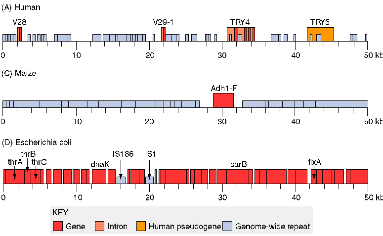
A genome is the total of all genetic sequence in an organism. See The Human Genome Project. The genome of Escherichia coli contains 4.6 million base pairs, encoding 4,400 genes. The human genome contains 3 billion base pairs in the nucleus, but only 30,000 to 40,000 genes (estimated), taking up 3% of the sequence. The rest includes regulator regions and large stretches of repetitive sequence of unknown function. The entire genome has been sequenced for several microbes as well as a number of animals and plants. For example:
|
What might the chimps have to say about it? Chromosome Structure Prokaryotes
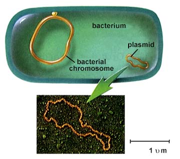 From Bacterial Diversity The bacterial DNA is packaged in loops back and forth. The bundled DNA is called the nucleoid. It concentrates the DNA in part of the cell, but it is not separated by a nuclear membrane (as in eukaryotes.) The DNA does form loops back and forth to a protein core, attached to the cell wall. 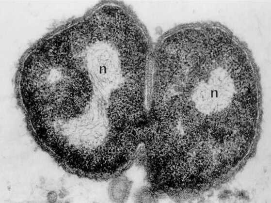 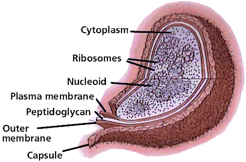 From Bacterial Diversity Eukaryotic cells contain their DNA within the nuclear membrane.
The DNA is wrapped around the histone core of eight protein subunits, forming the nucleosome. The nucleosome is clamped by histone H1. About 200 base pairs (bp) of DNA coil around one histone. The coil "untwists" so as to generate one negative superturn per nucleosome. 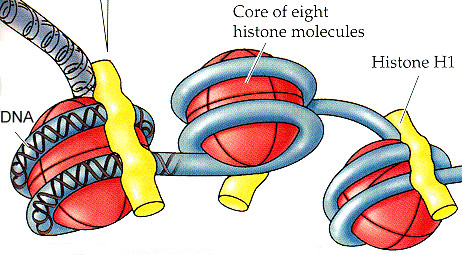 Life, the Science of Biology, by Purves, Orians, & Heller, 5th ed., 1997 Click on image to see molecular structure from Protein Data Bank (pdb 1aoi) This
form of DNA is active chromatin;
it can be "expressed" (transcribed and translated) to make RNA and
proteins
(Week 4, 5).
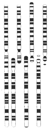 Life, the Science of Biology, by Purves, Orians, & Heller, 5th ed., 1997 The ribbon forms a coil, which then loops back and forth attached to a nuclear matrix -- similar to the protein core of bacteria, but greatly extended. During mitosis, several more layers of coiling result in fully condensed chromatin (see textbook Ch. 9). |
| In
mitosis,
the chromosomes appear as the thick rod-shaped bodies which can be
stained
and visualized under light microscopy.
The modern
way to visualize
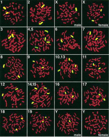
condensed chromosomes is by FISH --
fluorescence
in situ hybridization. In this
method,
fluorescent antibody-tagged DNA probes hybridize to their complementary
sequences in the chromosomes. By using FISH probes with different
colored fluorophores, one can color each human chromosome
independently,
and thus identify all 23 chromosomes. This is called
chromosome
painting.
|
 |