Biology Dept Kenyon College |
Overview of Development |

|
Biology Dept Kenyon College |
Overview of Development |
|
| Plant
development:
Embryogenesis
Invertebrate development: Drosophila Vertebrate development: Frogs and humans Invertebrate development: Drosophila Drosophila
and human development use closely related genes working in highly
conserved
regulatory networks. Unlike humans, Drosophila is subject to
easy
genetic manipulation. As a result, most of what we know about the
molecular
basis of animal development has come from studies of model systems such
as Drosophila.
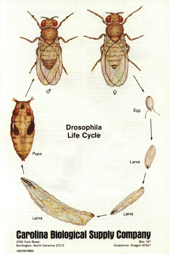
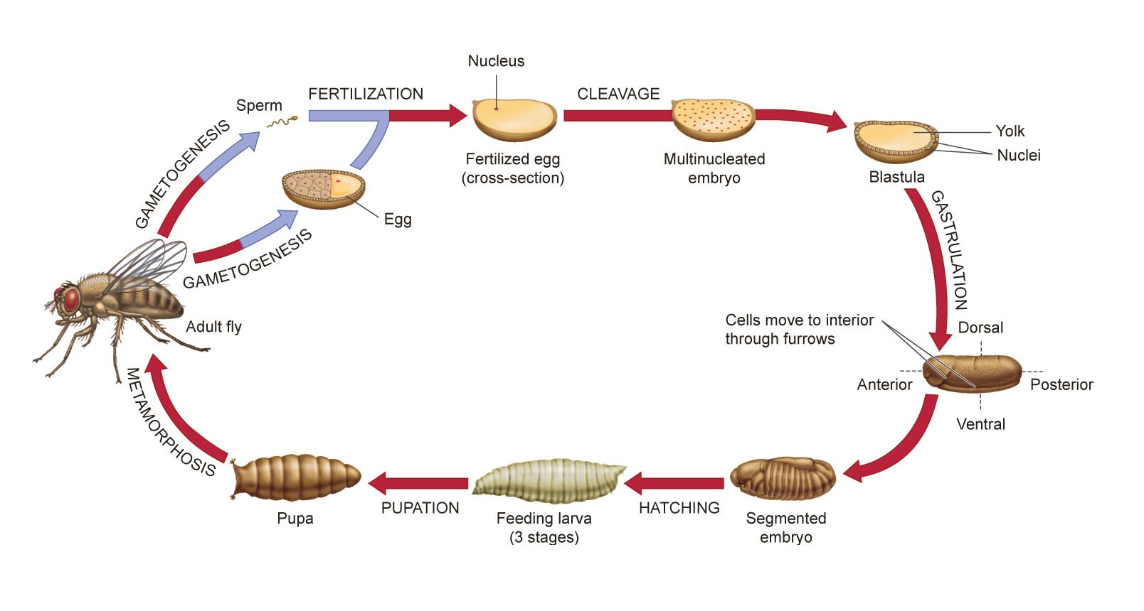
The Drosophila life cycle consists of a number of stages: embryogenesis, three larval stages, a pupal stage, and (finally) the adult stage!
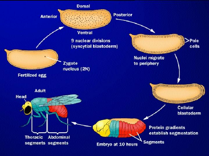
Embryogenesis in Drosophila Following
fertilization,
mitosis (nuclear division) begins. HOWEVER, cytokinesis (division of
the
cytoplasm) does not occur in the early Drosophila embryo,
resulting
in a multinucleate cell called a syncytium, or syncytial
blastoderm.
The common cytoplasm allows morphogen gradients to play a key role in
pattern
formation. At the tenth nuclear division, the nuclei migrate to the
periphery
of the embryo. At the thirteenth division, the 6000 or so nuclei are
partitioned
into separate cells. This stage is the cellular blastoderm.
Although
not yet evident, the major body axes and segment boundaries are
determined.
Subsequent development results in an embryo with morphologically
distinct
segments.
from LIFE: The Science of Biology, Purves et al, 1998 At the same time that segment patterns are being set up, cellular movements during the process of gastrulation result in the formation of the germ layers (ectoderm, mesoderm, and endoderm).
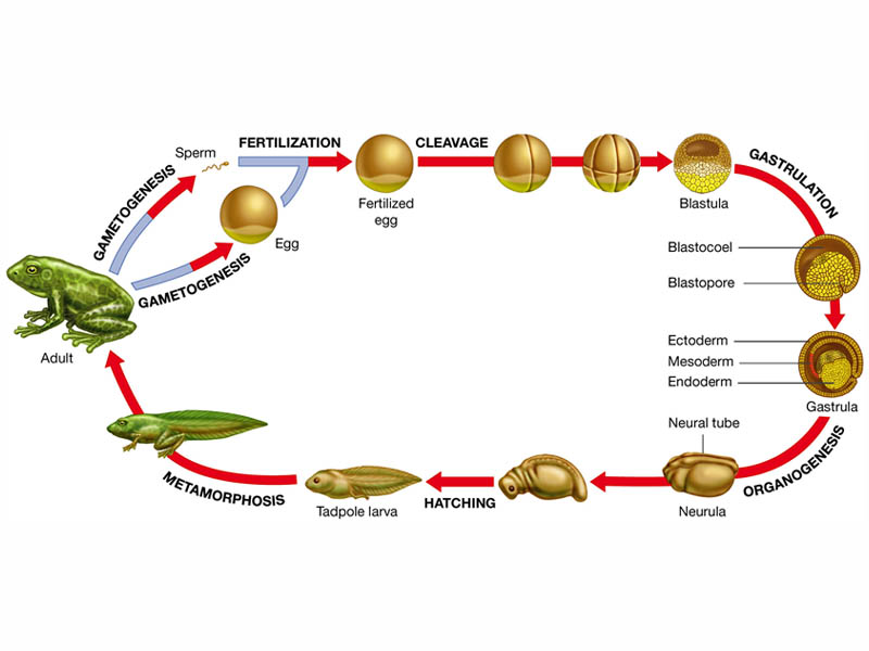
Vertebrate
development: Frogs and humans
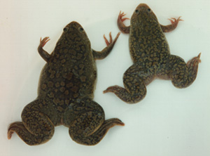
Although at first
glance
frog development looks quite different from that of Drosophila, the
same
stages of cleavage (cell division without cell growth) and gastrulation
(cell movements) are involved.
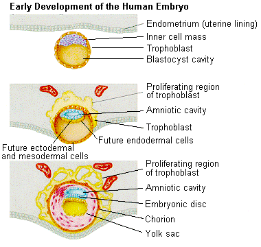
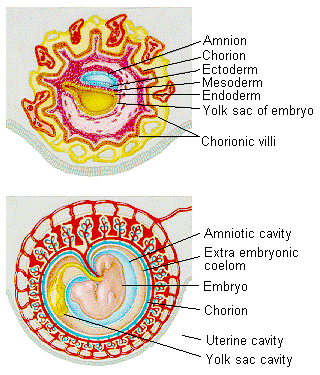
You can learn more about Frog embryology in Kimball's biology pages. Mammalian
development differs
from that of other vertebrates because embryonic development occurs
within
the mother, making the study of embryo development more difficult. The
developing embryo receives nutrients from the mother, and the eggs are
small and do not contain a large quantity of yolk - in contrast to
Drosophila
and frogs!
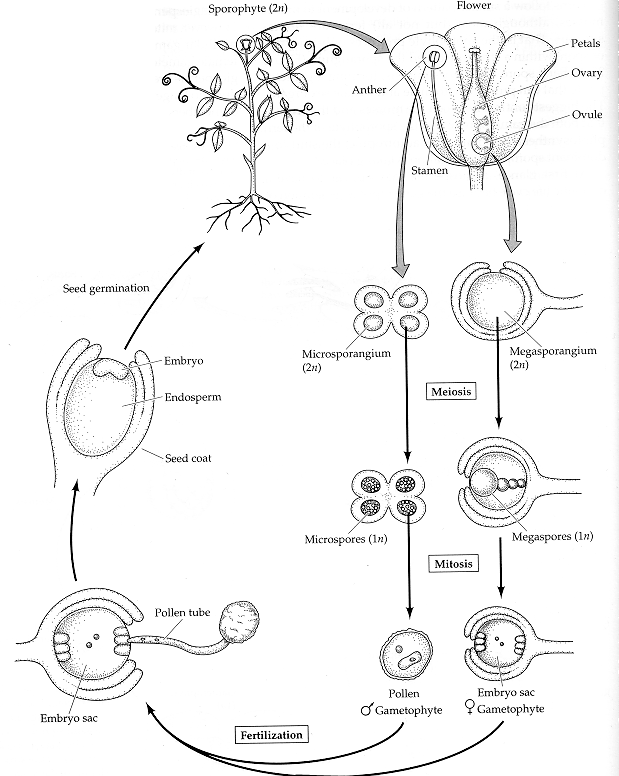
Plant development is different from animal development. Because plant cells have rigid cell walls, plant cells can't migrate. Therefore, plant shape is based on the rate and direction of cell division and cell elongation. Although plants develop three basic tissue systems (dermal, ground, and vascular), they don't rely on gastrulation to establish this layered system of tissues.The flowering plant (angiosperm) life cycle is shown below, and the fertilization process is shown in more detail after that.
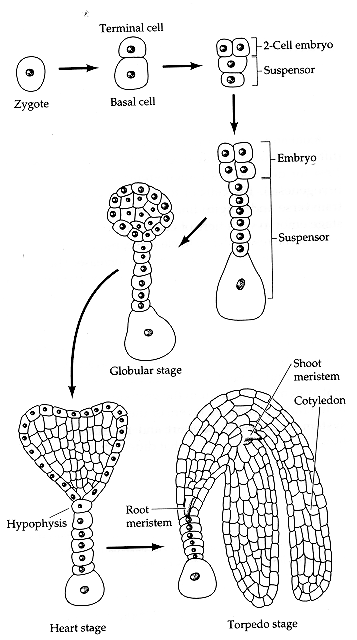
The egg cell and polar nuclei are contained within the embryo sac. The sperm nuclei are derived from the pollen grains. Double fertilization (fertilization by two sperm nuclei) results in a diploid zygote, which develops through embryogenesis into an adult plant, and a triploid endosperm, which provides nutrients to the developing embryo. Embryogenesis Plant embryogenesis begins with an asymmetric cell division, resulting in a smaller apical (terminal) cell and a larger basal cell. This first asymmetric division provides polarity to the embryo. Most of the plant embryo develops from the apical (terminal) cell. The suspensor develops from the basal cell. The suspensor anchors the embryo to the endosperm and serves as a nutrient conduit for the developing embryo. Further cell division leads to the globular stage. The three basic tissue systems (dermal, ground, and vascular) can be recognized at this point based on characteristic cell division patterns. The globular shape of the embryo is then lost as the cotyledons (embryonic leaves) begin to form. The formation of two cotyledons in dicots gives the embryo a heart-shaped appearance. In monocots, only a single cotyledon forms. Upright cotyledons can give the embryo a torpedo shape, and by this point the suspensor is degenerating and the shoot apical meristem and room apical meristem are established. These meristems will give rise to the adult structures of the plant upon germination. Further growth of the cotyledons results in the torpedo and walking-stick stages. At this point, embryogenesis is arrested, and the mature seed dessicates and remains dormant until germination.
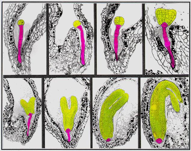
from Susan Singer In the following
images,
the descendants of the apical cell are shown in yellow, and the
descendants
of the basal cell are shown in pink.
A large amount information on cell division patterns and organogenesis during embryo development has been accumulated based on descriptive studies. However, in order to reveal the mechanisms underlying the pattern formation during plant embryogenesis, one needs to experimentally perturb this process. Two approaches, experimental embryology and genetic dissection, have been used for this purpose. Because plant embryos are not easily accessible (they are developing within the ovule of the maternal parent), experimental embryology has relied on somatic embryogenesis - formation of embryos from adult cells in tissue culture . However, this approach is problematic since a high proportion of abnormal embryos occur quite often in tissue culture. In the past
decade, many
scientists have been attempting to genetically dissect the mechanisms
underlying
plant embryo pattern formation. This approach relies on the isolation
and
characterization of mutants which are defective in this process,
primarily
using the model plant Arabidopsis thaliana.
from Detlef Weigel |