Human EMG

Muscles that control the hand originate on the medial and lateral surfaces of the humerus. They pass through the carpel tunnel and connect by a ligament to the bones of the fingers. Rest your arm on the table with your palm facing up. Alternately bend your fingers then tightly squeeze your hand and note what is happening to the muscles in your forearm. These muscles are flexors of the fingers. The extensors lie along the surface of the arm resting on the table. The finger flexors lie in several layers along the forearm and the tendons that attach them to the bones passes through the carpel tunnel, a space created by a broad ligament that lies near the wrist. It is often associated with carpel tunnel syndrome, a condition that occurs when the area through which the tendons pass into the hand becomes narrow and the medial nerve is compressed. Follow this link to a site created by J. Crimando at Gateway Community College if you would like to learn more about forearm anatomy.

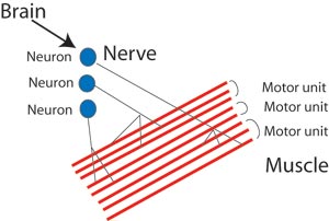
A simplified neural pathway containing 3 motor units.

To control the fingers the brain sends electrical signals that travel through individual neurons, stimulating the release of acetylcholine (ACh) from vesicles at the end of the neuron. ACh crosses the neuromuscular junction and stimulates muscle cells to contract.

  • A single neuron and the muscles cells it attaches to make up a motor unit.
  • The size of a motor unit depends on the function of the muscle and can contain from 10 to over 3000 muscle fibers.
  • Motor units in muscles that flex the fingers are small allowing for fine control.
  • Your brain determines the number of motor units in a muscle that contract as well as the rate that signals are sent to the motor units.
By placing electrical leads on the forearm it is possible to measure changes in electrical potential that are created on the surface of the skin when a muscle contracts. These changes are sent to a data acquisition unit that calculates the difference in electrical potential between the positive and negative electrodes and a record of the changes is graphed. This type of recording is known as an electromyogram (EMG).

Human forearm EMG recorded during flexion of the fingers.

Location of leads for recording an EMG from the flexor muscles of the forearm

Differences in the amplitude of the EMG recording on the left reflect differences in the number and size of active motor units. The brain uses sensory receptors in the muscles and tendons to determine the number of muscle units to recruit to perform a specific level of work

Human EMG recorded during squeezing a ball with increasing force.


In the trace on the left the brain is controlling the amount of force used to flex the fingers. Note that there is a small amount of electrical activity between the major pulses. This illustrates tonus, a constant state of slight contraction created by alternating periods of activation and rest in a few of the motor units that comprise the muscle. Tonus prepares the muscle for activity.

The increased amplitude seen in the trace resulted from recruiting additional motor units creating a graded response. If a muscle is functioning at a submaximal level the brain can cycle between motor units to keep the force relative constant. The motor units that relax can replenish their energy stores while those recruited perform the work. If muscles are forced to function at a maximal level or for prolonged periods of time they will fatigue. Fatigue results when muscles use energy faster than it is replenished and waste products accumulate.

Cross-section of Xenopus laevis gastrocnemius muscle 64X.
Each circle represents an individual muscle fiber.
Skeletal muscles of vertebrates contain several fiber types. These affect how quickly a muscle can repsond and how quickly it fatigues. Josh Mitchell '08' and Michael Northcutt '08' studied the distribution of fast and slow twitch fibers in the Northern Leopard Frog (Rana pipiens) and the African Clawed Toad (Xenopus laevis). They discovered that there were four different fiber types in the African Clawed Toad based on the intensity of succinate dehydrogenase staining. Differences in staining reflects differences in the number of mitochondria in the muscles. Fast fibers have fewer mitochondria than slow fibers and rely on anaerobic metabolism for energy rather than respiration. They respond rapidly and fatigue rapidly. In the figure on the left the fast fibers are lightly stained and larger than the slow fibers. If you would like to learn more about muscle fibers in amphibians take a look at the poster outside Higley 109.
QUESTIONS