Biology Dept Kenyon College |
DNA Structure and Mendelian Inheritance |

|
Biology Dept Kenyon College |
DNA Structure and Mendelian Inheritance |
|
DNA Structure Note: Much of this modified from the MIT Hypertextbook. Deoxyribonucleic acid (DNA) was first identified in 1868 by Friedrich Miescher, a Swiss biologist, in the nuclei of pus cells obtained from discarded surgical bandages. The substance he found contained an acidic part, nucleic acid, and a basic (alkaline) part, which we now know to be histone proteins, which bind to the nucleic acid. Which component was the genetic material? Many scientists were sure that it was protein. After all, protein had so many subunits (20 amino acids) that it seemed obvious that there existed within protein the possibility for much more diversity in expressing the genetic code than in DNA, which only has 4 subunits. Each subunit is identical except for the base: Learn these structures. Click here for Quiz! The Transforming Principle - DNA Might be the Genetic Material In 1943, Oswald Avery, Colin Macleod, and Maclyn McCarty, at the Rockefeller Institute, discovered that different strains of the bacterium Strepotococcus pneumonae could have different effects on a mouse. One virulent strain could kill an injected mouse, and another avirulent strain had no effect. When the virulent strain was heat-killed and injected into mice, there was no effect. But when a heat-killed virulent strain was coinjected with the avirulent strain, the mice died. What transforming principle was the dead virulent strain giving to the avirulent strain to make it lethal? This
phenomenon of
transformation,
the
uptake of DNA and incorporation into a genome, is now commonly
performed
in biotechnology..
Chargaff - Nucleotide Content in DNA In 1950, Erwin Chargaff at Columbia University discovered that no matter what tissue from an animal he looked at, the percentage content of each of the four nucleotides was the same, though the percentages could vary from species to species. In all animals:
%G = %C
The
significance of
these results was overlooked for three years, but they were crucial to
elucidating the structure of DNA.
Watson and Crick - The Double Helix In late 1953, James Watson and Francis Crick presented a model of the structure of DNA (see their paper in Nature.) It was already known from chemical studies that DNA was a polymer of nucleotide (sugar, base and phosphate) units. X-ray crytallographic data obtained by Rosalind Franklin, combined with the previous results from Chargaff and the chemists, were fitted together by Watson and Crick, who "borrowed" the data from Franklin's grant proposal. After several false starts, including the wrong tautomeric forms of the bases, they devised this model:
Under most cellular conditions, this double-stranded DNA molecule will coil naturally into a B-form helix, with one turn per 10.4 base pairs. However other structures are possible (see below). Each strand of the DNA is composed of nucleotides: The nucleotides form base pairs:  MIT Hypertextbook. Adenine
pairs with Thymine because they
make
two
hydrogen bonds.
The stacked base pairs form a major groove and a minor groove. Different regulatory proteins will bind to the major or minor groove. See Space-filling Model. Each base attaches to a phosphate at its 3' OH, and its 5'OH. The 2' carbon position has no OH; hence the "deoxy" part of DNA. The lack of 2' OH greatly stabilizes DNA, compared to RNA, because it prevents the intramolecular hydrolysis of phosphate linkages. The base
pairs "stack"
together like rungs on a ladder, because of favorable interactions
between
the pi orbitals extending out of the heteroaromatic ring structure of
each
base.
Helical forms of DNA The
structure of B-form
DNA helix was first determined by X-ray analysis of crystalized
molecules.
Certain regulatory sites within cells appear to have DNA sequence that takes a non-standard form, sometimes assisted by a protein. Moreover, DNA technologists are making use of unusual DNA properties to construct genetic medicines. Genetic medicines are pieces of artificial DNA that can hybridize to a region of the genome and turn off transcription of genes, such as a cancer gene. Stability
of DNA
However, in
water solution
certain chemical conditions can destabilize DNA.
Supercoiling In nearly all living cells, DNA contains negative superturns. This means it is "underwound," like a piece of yarn that has been twisted in the opposite direction that the multiple strands are wound. This is called negative supercoiling. Negative supercoiling may assist replication and transcription of DNA by lowering the energy needed to melt the helix. See the molecule Topoisomerase. In bacteria, negative superturns are maintained by the closed circular structure of the chromosome: It is impossible to unwind the superturns. In eukaryotes, negative superturns are maintained by the winding of the DNA helix around histone proteins. Problems. 1. Early life simulation experiments show that the base adenine would have formed spontaneously out of hydrogen cyanide, on the anaerobic early Earth. Show how five molecules of HCN can fit together to form exactly one molecule of adenine. 2. What kind of charge is on most of the proteins that take up 60% of the chromosome? Why? 3. If chemical analysis of a genome reveals 23% guanine, what are the percentages of the other three bases-- A, T, and C? 4. If a certain DNA site needs to come apart easily, for regulatory functions, what kind of base pairs are likely to be favored at that site? 5. Suppose an aromatic molecule with lots of pi orbitals could insert between two base pairs like a sandwich. What would happen as enzymes "read" the DNA information? 6. Some Archaea (microbes of the third Kingdom of organisms) living at extremely high temperature and pressure have positively supercoiled DNA. Why? DNA Replication DNA replicates semiconservatively. Replication starts by opening of the DNA helix at a particular sequence called an origin of replication (ori). Bacteria, even during logarithmic growth, have ONE map position where DNA can originate replication. Eukaryotes have MANY origins of replication, all of which run concurrently. In either case, each origin of replication runs bidirectionally, with TWO replicating forks.  Bidirectional semiconservative replication can be demonstrated by observing DNA from cells replicating in the presence of radiolabeled nucleotides. Both sides of the dividing DNA will be labeled. What would the above diagrams look like, if the replicating DNA were radiolabeled? Molecular Steps of DNA Replication Replication of DNA is mediated by enzymes and binding proteins. One crucial function is to unwind the helix, enabling it to "unzip" exposing bases to pair with the growing strand. How can the helix be unwound without falling apart? To see an example, view topoisomerase I. DNA replication must be fast and accurate. To follow the step by step process, click the image: 
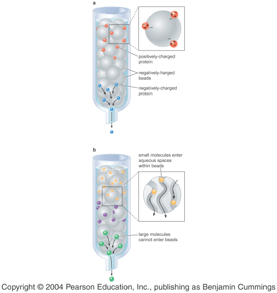
DNA polymerases and other factors involved in the process were initially discovered through protein purification efforts. Key to this technique is liquid chromatography.
Assumptions of Mendelian Inheritance
Mendelian inheritance Use Flowers tutorial: p:\data\biology\biol14\tutorial\flowers.exe Know these terms:
Mendelian Genetics Practice from a course at MIT. Virtual Fly : Breed your own fruit flies. Other links: Horse Genetics: describes interesting horse phenotypes and genotypes, crosses and results Online Mendelian Inheritance in Man: a professional physician’s reference on inherited diseases Alleles: What are they? An allele is a particular version of a given DNA sequence. "Allele" is a relative term, implying more than one possible version or copy, like different editions of a book. Like editions of a book, all existing alleles result from a process of change, either gradual or drastic change. Examples: Allele 1
Allele 2
Allele 3
Allele 4
Notice that
there can
be more than two possible alleles for a given gene locus (but
only
two at a time, in a given diploid individual.)
Natural and "artificial" alleles
Alleles can be observed as DNA polymorphisms, using restriction digest and gel electrophoresis (see Week 7). An allele may be linked to an inherited disease--a clue as to the gene locus defective in the disease. Which of the four alleles (M1-M4) is linked to this disease? Is the disease likely to be dominant or recessive?
For a Kenyon student's report on an inherited disease in her family, see Tuberous Sclerosis Complex. Alleles confer traits, by expressing gene products, which are either mRNA and protein, or a functional RNA. But how they determine a "visible trait" is not simple. Consider this: Fruit
fly eyes have two pigments, brown and scarlet. Normal flies make
In practice, the most common "new" alleles (arising out of mutation) are often named for a phenotype resulting from the absence of their gene product. Thus the allele for a gene producing scarlet pigment is named "brown" for the brown eye in the absence of scarlet pigment. Consider
albinism,
or loss of pigmentation, a very common phenotype observed in many
species
of animals and plants. Alleles can confer loss of pigmentation in
two different ways:
The reason
Mendelian
inheritance can be seen to "work" is that in many cases we can hold all
the above factors constant, for a given genotype (genes
affecting
a trait) and a given phenotype (appearance of trait).
Other ways alleles can work (still at a SINGLE gene locus)
Pleiotropy. One allele (or pair of recessive alleles) at one gene locus can result in many diverse effects throughout the body.
Dihybrid Cross -- or local link. Advanced Problem. Explain how to breed transgenic mice to create a mouse model for sickle-cell anemia.  Pászty et al, Science 1997 October 31; 278: 876-878. Sickle-cell disease is caused by a single base pair defect in human beta-globin.
For the model strain to exhibit sickle-cell pathology, the native mouse genes--all at separate loci--must be defective (null alleles.) We have a transgenic mouse strain, containing human Hb-alpha, Hb-beta-sickle on a transgene,Tg(Hu), inserted somewhere in the mouse genome (not at the mouse globin genes.) But the mouse still has its own genes producingalpha and beta globin. To construct this strain, the transgenic strain was intercrossed with a mouse strain heterozygous for null alleles for alpha and beta globin. Tg(Hu)
Mouse-alpha-Hb
Mouse-beta-Hb
X
-- Mouse-alpha-Hb
Mouse-beta-Hb
How many
generations
would you need to cross?
Tg(Hu)
----
----
What would
the researchers
have to do to create a similar model with exclusively normal human
blood?
Why would this be important in order to use the model? (For
further
interest, read Ryan
et al, 1997.)
|
